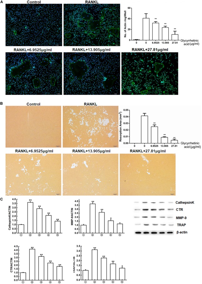
FIGURE 2.

18β-GA inhibits osteoclasts function in vitro. (A) Actin ring structures of osteoclasts and quantification of the actin ring. (B) Pits formation assay of osteoclasts and quantification of pits area.  RAW264.7 cells;
  RAW264.7 cells induced with M-CSF, RANKL, and PBS;
  RAW264.7 cells induced with M-CSF, RANKL and treated with 6.9525 μg/ml 18β-GA;
  RAW264.7 cells induced with M-CSF, RANKL and treated with 13.905 μg/ml 18β-GA;
  RAW264.7 cells induced with M-CSF, RANKL and treated with 27.81 μg/ml 18β-GA. (C) Western blot and optical density analysis of the expression of Cathepsin K, CTR, MMP-9, and TRAP with β-actin as reference (∗P < 0.05, ∗∗P < 0.01 versus
  ).
).
