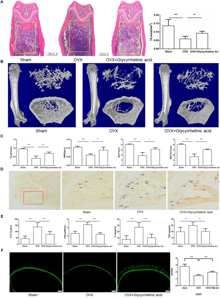
FIGURE 7.

18β-GA ameliorates ovariectomy-induced bone loss in vivo. (A) Representative HE staining of distal femoral sections and quantification of trabecular area from each group 6 weeks after the operation. (B) Micro CT analysis of the distal femur from sham, OVX, and OVX + 18β-GA group. (C) Calculations of trabecular number (Tb.N), bone mineral density (BMD), bone surface area/total value (BS/TV) and bone value/total value (BV/TV). (D) Representative TRAP-stained histologic distal femur sections from sham, OVX and OVX + 18β-GA group. (E) Serum IL-6, TNF-α, TRAcp5b, and CTX-1 were examined. (F) Calcein double labeling with a 7-day interval (Scale bar: 100 μm). Mineral apposition rate (MAR) is significantly decreased in OVX mice and reversed by 18β-GA (∗P < 0.05, ∗∗P < 0.01, ∗∗∗P < 0.001).
