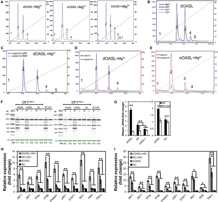
Figure 3.
Duck and ostrich OASL proteins synthesize 2–5A to activate and enhance the OAS/RNase L pathway degrading cellular and viral RNA. The 2–5A synthetase reaction was treated with alkaline phosphatase and separated using a Mono Q column. hOAS1 was a positive control for 2–5A activity. Numbers in the elution profiles of A–E represent adenylate, pppApA, pppA(pA)2, pppA(pA)3, pppA(pA)4, pppA(pA)5, pppA(pA)6, respectively. NC is chicken OASL-deficient DF1 cell expressing empty vector. WT is parent DF1 cell. “RIN” is the RNA integrity number. Gene expressions were calculated relative mRNA level to that of GAPDH and presented as fold change against the corresponding of NC without CK/0513 infection (two-tailed Student’s t-test, n = 3). The data are expressed as the mean ± SD. *P < 0.05; **P < 0.01. (A–E) 2–5As produced by dOASL or oOASL with pIC (A), dOASL with different divalent cations (B), dOASL with HMW or LMW (C), dOASL with pAT (D), oOASL with pAT (E). (F) rRNA cleavage induced by dOASL or oOASL in DF1OASL−/− cells transfected with pIC (5 µg/mL) for 4 h or infected with CK/0513 (multiplicity of infection = 1) for 18 h. (G–I) dOASL and oOASL significantly increased the expression of RNase L (G) and 10 genes (H,I) related to IFN signaling after infection with CK/0513 virus in DF1OASL−/− cells, respectively.

