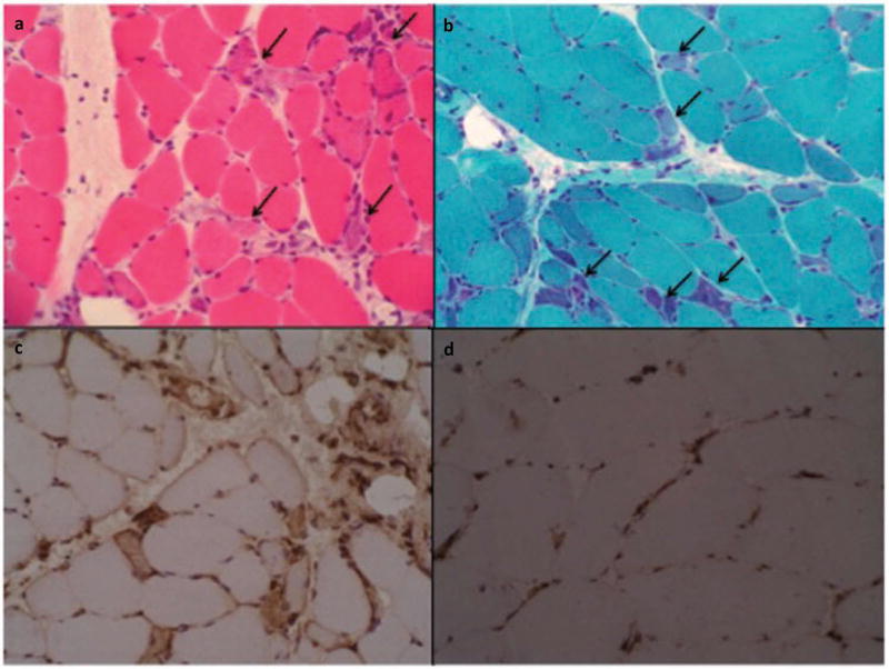
Figure 1.
Immune-mediated necrotizing myopathy. Necrotic muscle fiber cells (arrows). (a) Hematoxylin-eosin, (b) Numerous regenerating fibers are seen (arrows). Masson Trichrome. (c) Universal sarcolemmal MHC class I positive (stronger in regenerating cells). (d) MHC class I negative control (positivity only in endothelial cells). All samples are frozen tissue. Published with permission of Elsevier. Original source: Alvarado Cárdenas et al. Med Clin (Barc) [63]. Copyright © 2015 Elsevier España, S.L.U. All rights reserved.

