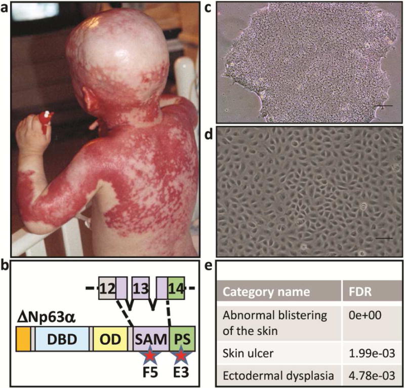
Figure 1. Generating iPSC and iPSC-derived keratinocytes from AEC patient skin.

(a) Patient affected by AEC exhibiting severe skin erosions. The patient and his parents consented to the use of this image in this publication. (b) TP63 mutations in AEC patients occur mainly in exons 13-14, encoding putative protein-protein interaction domains. Approximate location of mutations in the AEC patient cells (F5 and E3) used in this manuscript are indicated by stars. The protein schematic shown is of ∆Np63α, the predominantly expressed TP63 isoform in human keratinocytes. DBD: DNA binding domain, OD: oligomerization domain, SAM: sterile alpha motif, PS: post-SAM domain. Phase contrast images of (c) human iPSC colony and (d) iPSC-derived keratinocytes. (e) Disease pathways associated with the TP63 mutations in E3 and F5 iPSC-K as determined by a Human Phenotype Ontology analysis of our RNAseq data (FDR; false discovery rate).
