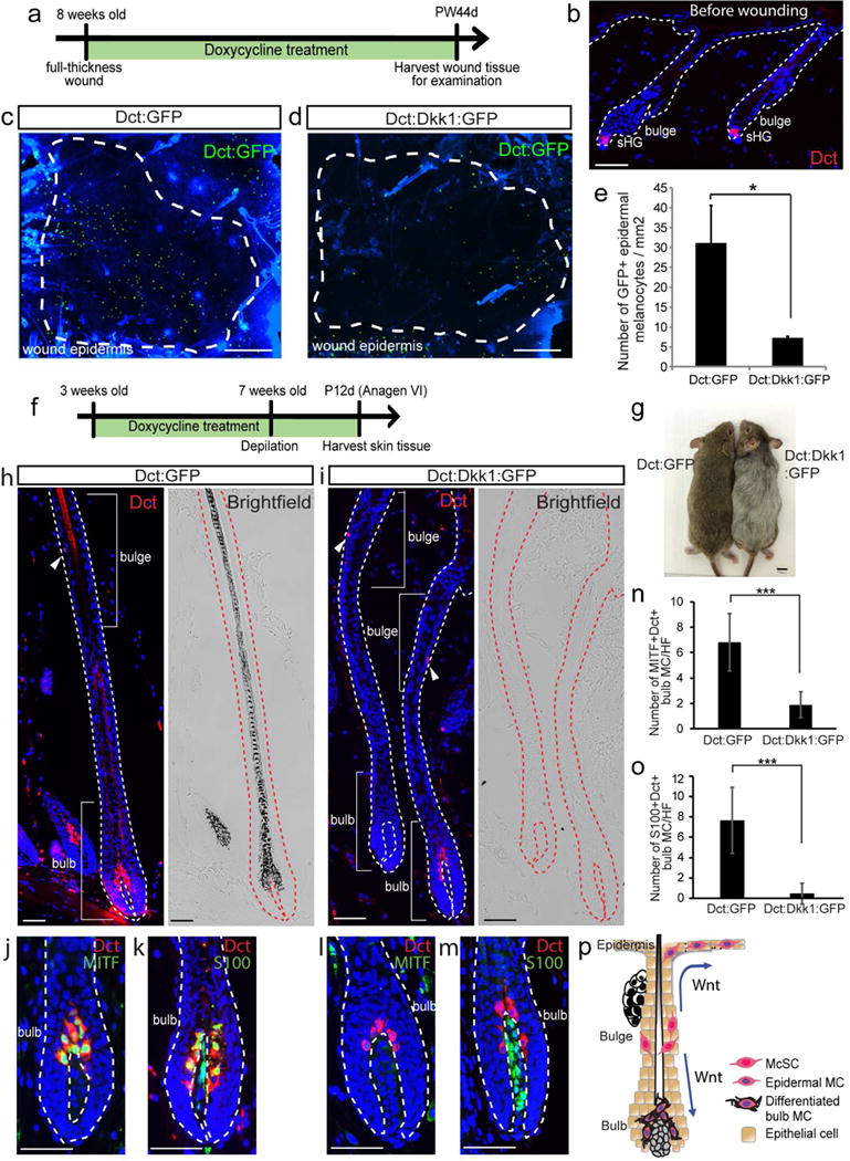
Figure 1. Overexpression of Dkk1 in melanocytes inhibits the wound-induced generation of epidermal melanocytes.

(a-e) Dct-rtTA; tetO-Dkk1; tetO-H2B-GFP (Dct:Dkk1:GFP) and Dct-rtTA; tetO-H2B-GFP (Dct:GFP) mice were wounded and treated with doxycycline for 44 days. (a) Experimental scheme. (b) Immunofluorescence of Dct before wounding. (c, d) Dct:GFP signals in whole mount wound epidermis at PW44d. (e) Quantification of (c-d). (f-o) Dct:Dkk1:GFP and Dct:GFP mice were treated with doxycycline from 3 weeks old and depilated at 7 weeks old. Tissues were harvested at P12d (12 days after depilation). (f) Experimental scheme. (g) Image of mouse at P12d. (h-m) Immunofluorescence of indicated markers and corresponding brightfield images. (n-o) Quantification of (j-m). S100+/Dct− cells in (k, m) are dermal papilla cells. (p) Schematic summary. Dashed lines in (c-d): boundary between wound and intact areas. Dashed lines in (b, h-m) boundary of epithelium and dermis. Arrowheads: melanocyte stem cell (McSC). PW: post wound. MC: melanocytes. HF: hair follicle. sHG: secondary hair germ. Data are represented as mean ± SEM; * p<0.05, *** p<0.001. Scale bar: 50um in (b, h-m), 500um in (c-d), 1cm in (g).
