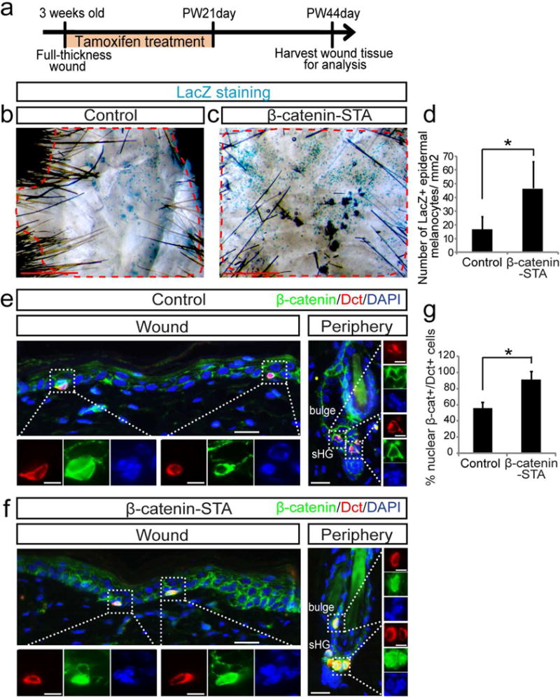
Figure 2. Overexpression of β-catenin promotes the generation of epidermal melanocytes following wounding.

Tyr-CreER; β-catenin fl(ex3)/+; Dct-LacZ (β-catenin-STA) and Dct-LacZ control mice were wounded and treated with tamoxifen for 21 days. Wound tissues were harvested at PW44day. (a) Experimental scheme. (b, c) β-galactosidase staining of whole mount wound epidermis. (d) Quantification of (b, c). (e, f) Immunofluorescence of indicated markers in wound center and hair follicles in wound periphery. (g) Quantification of the percentage of nuclear β-catenin+ melanocytes in wound epidermis of (e, f). Dashed lines outline the boundary between wound and intact areas. Dotted boxes outline magnified regions in separate fluorescent channels. PW: post wound. Data are represented as mean ± SEM; * p<0.05. Scale bar: 1000um in (b, c), 25um in (e, f) and 10um in magnified images in (e, f).
