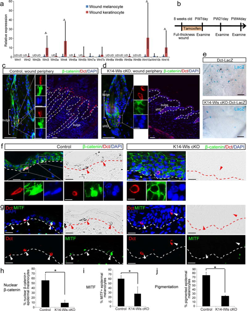
Figure 3. Wnt ligands provided by epithelial cells are required for Wnt activation and differentiation of epidermal melanocytes.

(a) Wnt ligand qPCR of epidermal melanocytes and keratinocytes isolated from wound epidermis 17 days after wounding. UD: undetected. (b-j) K14-CreER; Wls fl/fl (K14-Wls cKO) or K14-CreER; Wls fl/fl ; Dct-LacZ (K14-Wls cKO-Dct-LacZ) mice and control littermates were wounded and immediately treated with tamoxifen for 7 days. Wound tissues were harvested at indicated time points. (b) Experimental scheme. Immunofluorescence of indicated markers and corresponding brightfield images at PW7day (c, d) and PW21day (f, g). (h-j) Quantification of (f, g). (e) β-galactosidase staining of whole mount wound tissue at PW44day. Dashed lines outline the boundary of epithelium and dermis. Dotted boxes outline magnified regions in separate fluorescent channels. Arrowheads point to epidermal melanocytes. PW: post wound. Data are represented as mean ± SEM; * p<0.05. Scale bar: 50um in (c-e), 25um in (f, g), 10um in magnified images in (c, d, f).
