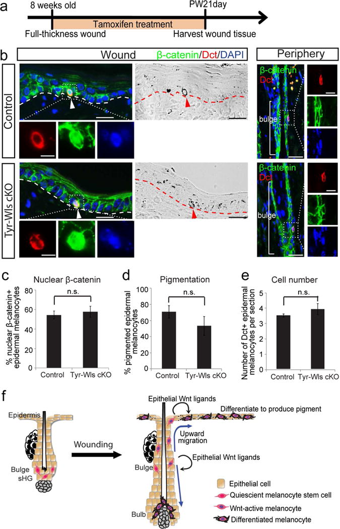
Figure 4. Melanocytes do not produce Wnt ligands to drive their Wnt activation and differentiation.

(a-e) Tyr-CreER; Wls fl/fl (Tyr-Wls cKO) mice and control littermates were wounded and immediately treated with tamoxifen for 21 days. Wound tissues were harvested 21 days after wounding. (a) Experimental scheme. (b) Immunofluorescence of indicated markers and corresponding brightfield images in wound center and hair follicles in wound periphery. (c-e) Quantification of (b). (f) Schematic model: Upon wounding, epithelial cells generate Wnt ligands to drive the Wnt activation and upward migration of quiescent McSCs. Once in the epidermis, Wnt ligands from epithelial cells continue to induce Wnt activation in epidermal melanocytes, which differentiate and produce pigment. Wnt activation is also required for McSCs to generate hair bulb melanocytes. Dashed lines outline the boundary of epithelium and dermis. Dotted boxes outline magnified regions in separate fluorescent channels. Arrowheads: epidermal melanocytes. PW: post wound. Data are represented as mean ± SEM; n.s.: not significant. Scale bar: 25um and 10um in magnified images.
