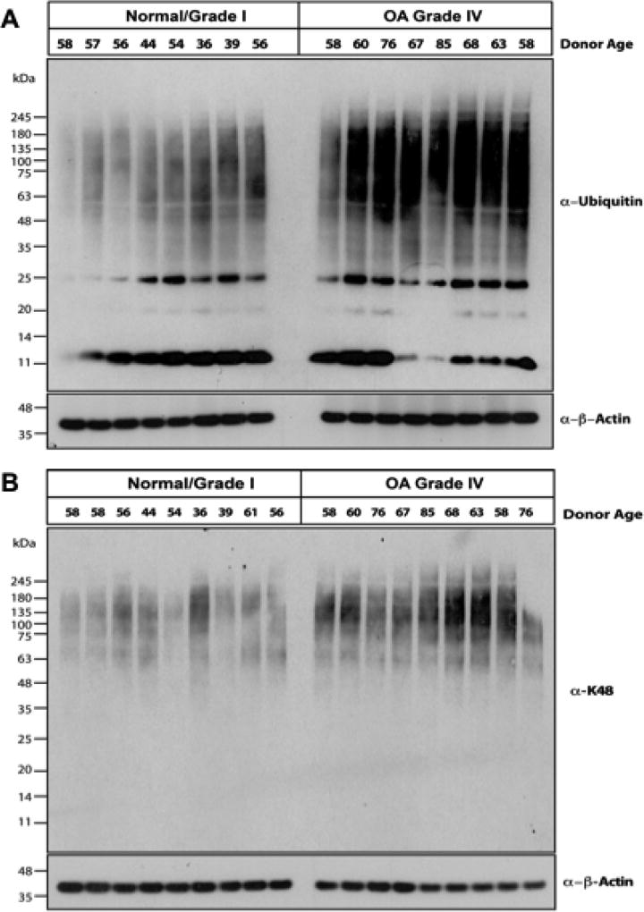
Figure 1. Increased ubiquitin and polyubiquitinated substrate levels in human knee OA chondrocytes.
Equal aliquots (10 µg) of lysates from normal (Grade 0/Grade I) and Grade IV OA human articular chondrocytes were used for 4–12% SDS-PAGE/Western blot analysis, employing antibodies directed against ubiquitin (Ub, n=16) (A) and K48-linked ubiquitin (n=18) (B). Equal protein loading was also confirmed by blotting for β-actin.

