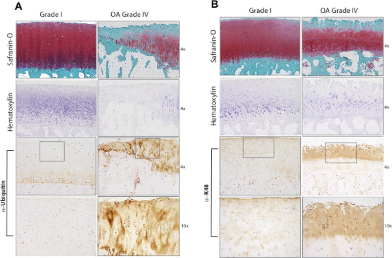
Figure 2. Increased levels of ubiquitin and polyubiquitinated substrates in human knee OA cartilages in situ.
Immunohistochemistry of paraffin embedded sections from grade IV OA and normal human knee articular cartilages employed antibodies specific for ubiquitin (Ub) (A) and K48-linked ubiquitin (K48) (B). Safranin-O, and hematoxylin cell staining are shown at 4× magnification. For IHC, 4× and 10× magnification results are shown, with the third panels from the top showing a boxed area magnified in the panels immediately below. Representative of 12 separate normal and 12 separate OA donors.

