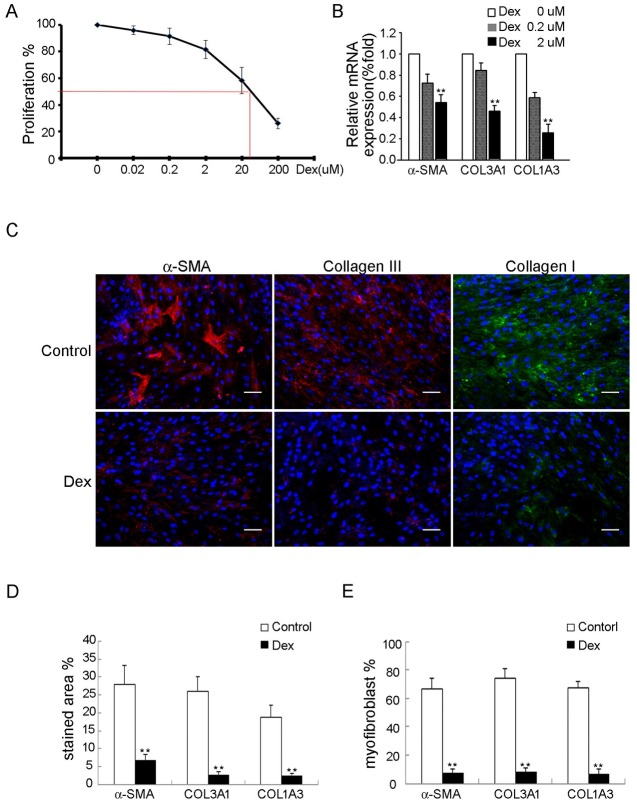
Fig 1. Dexamethasone inhibits in vitro myofibroblastic differentiation of FSCs.
(A) FSCs seeded at 2000 cells/well in 96-well plates were treated with dexamethasone at the indicated concentrations for 7 days. The IC50-value was measured. (B) mRNA expression of α-SMA, Col3A1 and Col1A3 was analyzed by quantitative RT-PCR after treatment with dexamethasone 0, 0.2 and 2 uM for 14 days. (C) Immunofluorescence staining for α-SMA (red), types III (red) and I collagen (green) after treated with either dexamethasone 2uM or 0 uM (control) for 14 days. Bars = 50 μm. (D) The percentages of stained areas. (E) The percentages of myofibroblasts. Data are shown as mean ± SD (n = 3). Statistical significance is presented as **, p<0.01 compared with other groups. All experiments were repeated with FSCs isolated from three different donors.

