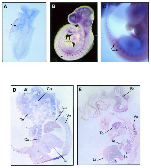
Figure 3.
Mouse and human Sox7 expressions as revealed by in situ hybridisations. Whole-mount in situ hybridisations showing expression of Sox7 in the mouse developing embryo. (A) Ventral view of an 8 d.p.c. embryo (4-somite stage) revealing Sox7 expression in the somites (black arrow) and the brain (white arrow). (B) At 9.5 d.p.c., Sox7 expression is observed in the intersomitic vessels, along with the smaller vessels in the trunk and the head region. (C) At 11.5 d.p.c., expression persists in the intersomitic vessels (black arrows). Section in situ hybridisations comparing expressions of Sox7 in mouse and human embryos. (D) Sagittal section of 17.5 d.p.c. mouse embryo showing generalised Sox7 expression in brain (Br), cochlea (Co), tongue (To), lung (Lu), liver (Li), cartilage (Ca) and vertebrae (Ve). (E) Sagittal section of an 8 week-old (C.S.19) human embryo showing expression in brain, tongue, heart, lung, liver and vertebrae. Embryo sections in (D) and (E) are counterstained with nuclear fast red.

