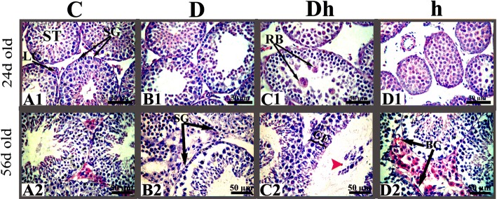
Fig. 3.
Histo-architectural changes in testicular cells during experimental diabetes mellitus and hypothyroidism in immature and prepubertal mice. Sloughed spermatids, marked with red arrow head shown inside the lumen of tubule of Dh animals (panel: C2). Abbreviations: Control (C), Diabetic (D), diabetic + hypothyroidism (Dh) and Hypothyroidism (h), Seminiferous tubule (ST), Blood cells (BC), Leydig cells (LC), Spermatogonia (SG), Germinal epithelium (GE),. Representative images were captured at 400× magnifications. The bars are 50 μm in size

