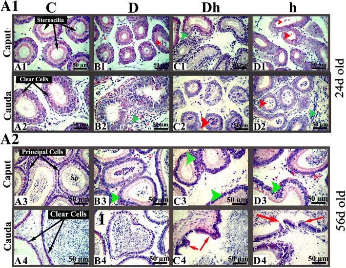
Fig. 4.
Hematoxylin and eosin stained digital images of the epididymis at different age levels of mice, showing proximal caput and distal cauda under the influence of diabetes and hypothyroidism compared with control. (A1) Epididymal sections of immature mice at the age of 24 days old. Most of the epididymal tubules of treated animals possess round bodies with exfoliated germ cells, marked with red arrow heads (panels B1, D1, C2 and D2). Some of the epididymal tubules of treated mice, depicts inflammatory infiltrations marked with green arrow heads (panels C1, B2 and D2). (A2) Epididymal sections of pre pubertal mice at the age of 56 days old presenting hyperplastic changes in principal cells of caput tubules are shown with green arrow heads (panels: B3, C3 and D3). Cribriform changes are marked with red arrows (pannels: C4 and D4). Abbreviations: Control (C), Diabetic (D), diabetic + hypothyroidism (Dh) and Hypothyroidism (h), (S) Spermatozoa and (I) Interstitium. The pictures were captured at magnification of 400× and the bars are 50 μm in size

