
Fig 6. Transcriptional profiling reveals TGF-β signaling components, BMP target genes, and hair follicle stem cell signature genes are downregulated in Bmp6 mutant tooth plates.
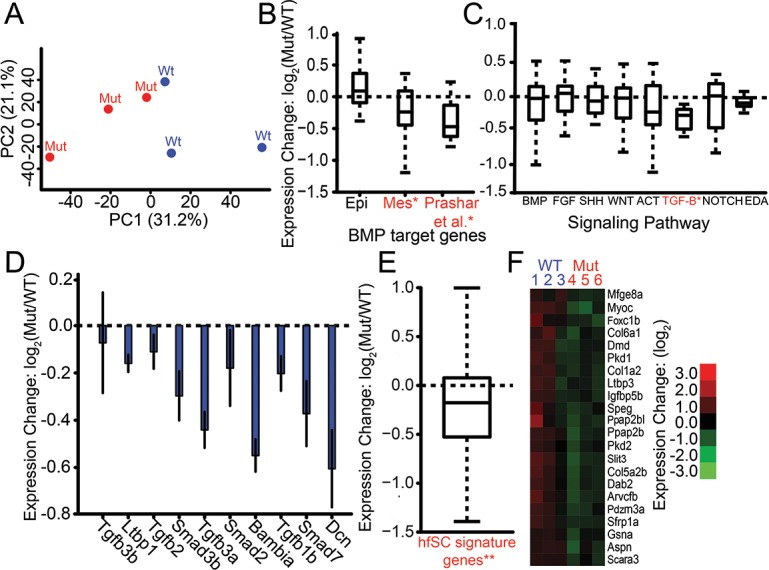
(A) Principal component analysis of genome-wide expression levels in late juvenile ventral pharyngeal tooth plate tissue by RNA-seq separates wild-type (Wt, blue) and Bmp6 mutants (Mut, red) along PC1. (B) BMP target genes in developing tooth epithelium and mesenchyme [8] were not affected (left bar), and significantly downregulated (P = 1.25 x 10⁻², middle bar), in the mutant, respectively. A set of BMP target genes [36] was significantly downregulated in mutants (P = 3.12 x 10⁻⁴, right bar). (C) Expression of ToothCODE signaling pathways. Homozygous mutant fish (Mut) had significantly lower TGF-β pathway expression compared to wild-type fish (WT) (P = 4.7 x 10⁻³). None of the other pathways showed significant differences. (D) Each of the ToothCODE TGF-β genes was downregulated in the mutant. Error bars are SE of the mean. (E) A previously described set of genes upregulated in the mouse hair follicle stem cell niche [46] was downregulated in Bmp6 mutants (P = 8.5 x 10⁻¹²). hfSC = hair follicle stem cells (F) Hair follicle stem cell signature genes showing significant downregulated expression in Bmp6 mutants. See S1 File for gene expression levels and gene sets. For B, C, and E, gene sets with significant expression differences between wild-type and mutant are listed in red and with an asterisk.