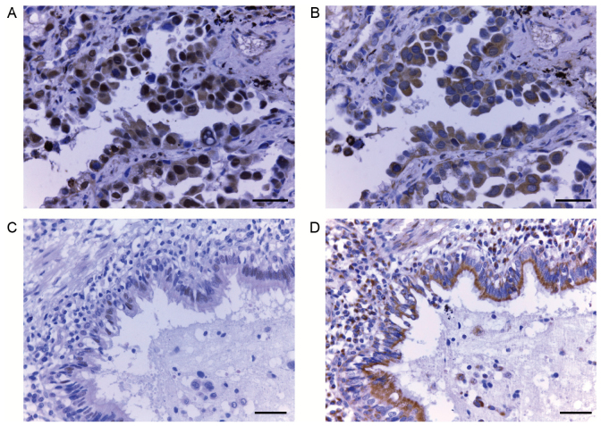
Figure 1.
Immunohistochemical staining for 14-3-3ζ and E-cadherin in lung adenocarcinoma tissues. (A) Representative picture of lung adenocarcinoma tissue showing positive 14-3-3ζ staining. Scale bar, 20 µm. (B) Representative picture of lung adenocarcinoma tissue showing positive E-cadherin staining. Scale bar, 20 µm. (C) Representative picture of lung normal tissue showing negative 14-3-3ζ staining. Scale bar, 50 µm. (D) Representative picture of lung normal tissue showing positive E-cadherin staining. Scale bar, 50 µm.

