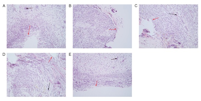
Figure 4.
Morphological changes of endometrium detected by hematoxylin and eosin staining in (A) control, (B) model, (C) EnSCs, (D) estrogen and (E) E+EnSCS groups. Black arrows indicate glands, red arrows indicate epithelial cells. Magnification, ×200. E+EnSCs, estrogen plus endometrial stem cells.

