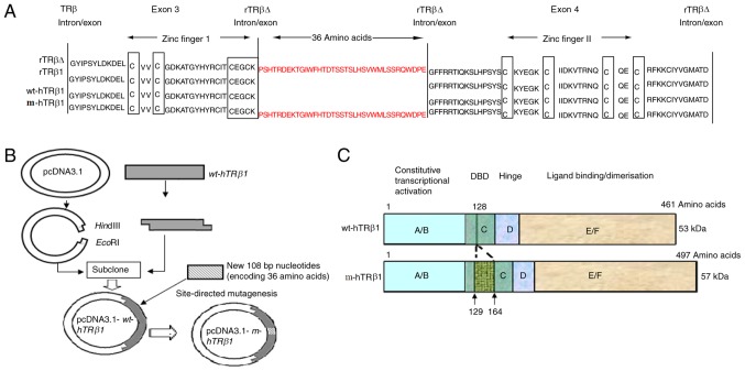
Figure 1.
Schematic of the wt-hTRβ1 and m-hTRβ1 protein structure and expression vector construction. (A) Amino acid sequences around the P box in the DBD of TRβs. The amino acid sequences are presented from exon 3–4 of TRβ. Homologous sequences are relative to the zinc finger motifs. A long gap is introduced for best alignment. The P box and the cysteines of the zinc fingers are boxed. The coding sequence of the 36 amino acids was introduced between exon 3 and 4 of wt-hTRβ. The mutant was termed m-hTRβ1 (B) Map of the pcDNA3.1-wt-hTRβ1 and pcDNA3.1-m-hTRβ1 plasmids, vector construction and the mutation are depicted. (C) Schematic diagram representing the protein structure of wt-hTRβ1 and m-hTRβ1. TRβ, thyroid hormone receptor β; wt-hTRβ1, wild-type human TRβ1; m-hTRβ1, modified human TRβ1; rTRβΔ, rat TRβ isoform; DBD, DNA-binding domain.

