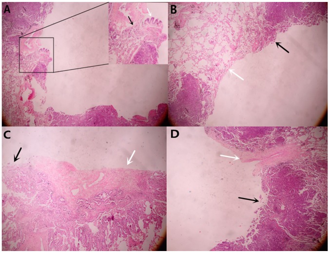
Figure 7.
(A) Tumor cells (black arrow) destroyed the bronchi wall (white arrow). (B) The cavity wall was covered with tumor cells in patients who underwent surgery. The black arrow represents the area of the wall covered with tumor cells and the white arrow represents the area of the wall not covered with tumor cells, which demonstrated that thin-wall cavity formation had been initiated prior to tumor cell covering the wall. (C) Hyperplastic fibrous tissue was observed inside the cyst. (D) The blood vessels (indicated by the white arrow) blocked the proliferation of tumor cells. Magnification, ×40.

