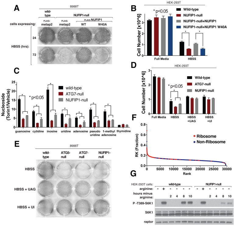
Fig. 5. NUFIP1 is important for cells to survive starvation.
(A) Loss of NUFIP1 or just its capacity to interact with LC3B impairs cell survival upon nutrient starvation. Wild-type or NUFIP1-null 8988T cells stably expressing the indicated proteins were deprived of nutrients by culturing them in Hank’s Balanced Salt Solution (HBSS); after the indicated times the surviving cells were stained and imaged.
(B) Loss of NUFIP1 or just its capacity to interact with LC3B impairs cell survival upon nutrient starvation. Wild-type or NUFIP1-null HEK-293T cells stably expressing the indicated proteins were deprived of nutrients by HBSS, and after 48 hours the number of surviving cells was quantified using cell counting. Values are normalized relative to cell numbers at the start of the starvation period and are mean +/− SD (*P<0.05; n=3).
(C) Loss of NUFIP1 or ATG7 inhibits the increase in nucleosides caused by mTOR inhibition. Data represent relative change in whole-cell concentrations of nucleosides in wild-type, ATG7-null, and NUFIP1-null HEK-293T cells treated with 250 nM Torin1 for 1 hour. Values are mean −/+ SEM (*P<0.05; n=3)
(D) Nucleoside supplementation rescues the survival defects of ATG7-null and NUFIP1-null HEK-293T cells. Indicated cells were deprived of nutrients by culturing in HBSS with or without the indicated nucleosides (2 mM each). After 48 hours, the number of surviving cells was quantified. Values were normalized relative to cell numbers at the start of the starvation period and are mean +/− SD (*P<0.05; n=3).
(E) Nucleoside supplementation rescues survival defect of ATG5-null, ATG7-null, and NUFIP1-null 8988T cells. Wild-type, ATG5-null, ATG7-null, or NUFIP1-null cells were deprived of nutrients by culturing in HBSS with or without the indicated nucleosides (2 mM each). After 48 hours the surviving cells were stained and imaged.
(F) Ribosomes are highly enriched for arginine and lysine. Protein sequences in the UniProt database (including isoforms) were ranked based on their fraction content of arginine and lysine. Ribosomal proteins are shown in red; all other proteins are shown in blue. Mitochondrial ribosomal proteins were not designated as ribosomal in this analysis.
(G) Loss of NUFIP1 suppresses the reactivation of mTORC1 that occurs after long-term arginine deprivation. Wild-type or NUFIP1 HEK-293T cells were deprived of arginine for 50 mins (first lanes of each set) or the indicated times and, where indicated, re-stimulated with arginine for 10 mins. Cell lysates were analyzed by immunoblotting for the levels and phosphorylation states of the indicated proteins.

