Fig. 5.
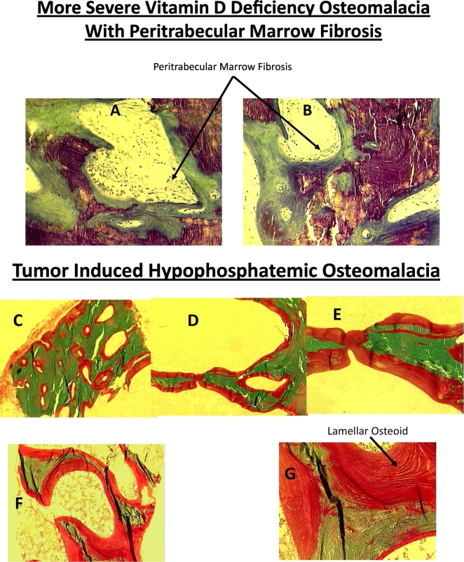
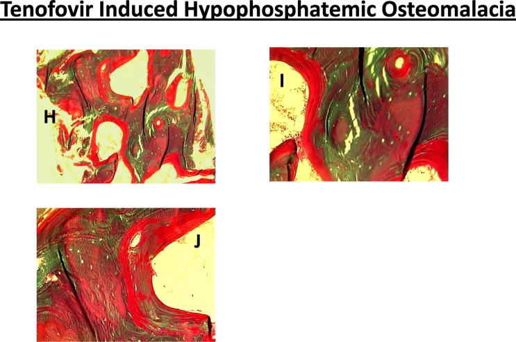
Representative bone biopsy photomicrographs in different types of osteomalacia. Increased osteoid thickness and surface (A & B) with marrow fibrosis (arrows) in a patient with severe vitamin D deficiency osteomalacia (Al-Shoha et al., 2009). In contrast, marrow fibrosis is not seen in tumor (C-G) or tenofovir induced (H-J) hypophosphatemic osteomalacia. Also, the osteoid thickness and surface (red color) is more extensive in both types of hypophosphatemic osteomalacia (C-J) compared to vitamin D deficiency osteomalacia (A & B). Finally, the osteoid is of lamellar type (arrow) rather than woven type as seen in fracture repair, Paget's disease of bone, osteitis fibrosa, all of which may also show increased osteoid. (For interpretation of the references to color in this figure legend, the reader is referred to the web version of this article.)


