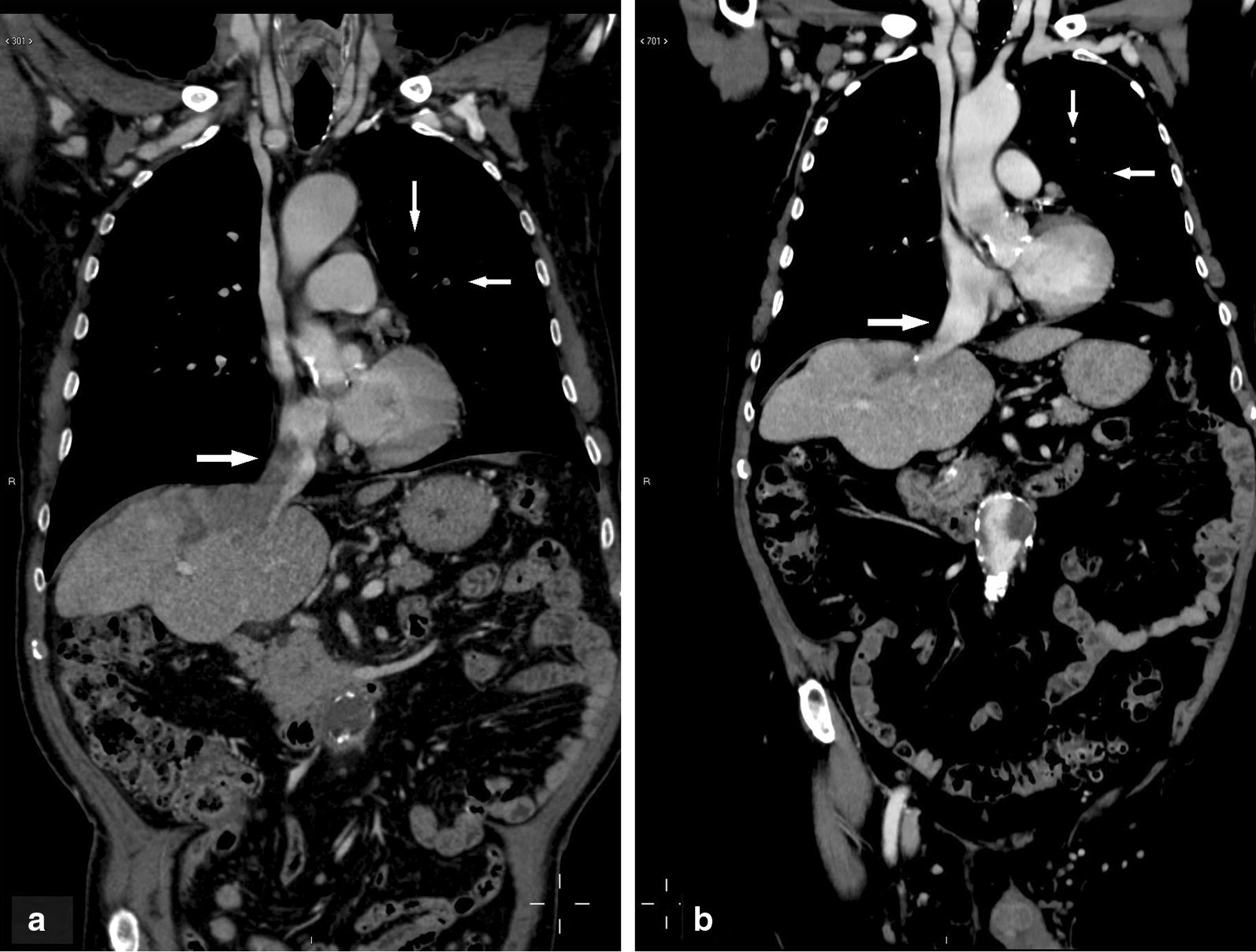
Fig. 3.

Lesions in case 3: a pre-treatment CT scan (coronal image of the portal fase). The image shows vascular invasion of the right and medium hepatic veins and of the inferior cava vein. Two neoplastic embolisms are present in the left lung. b Following 18 months of treatment with metronomic capecitabine, vascular vein invasion was resolved and pulmonary embolisms had disappeared
