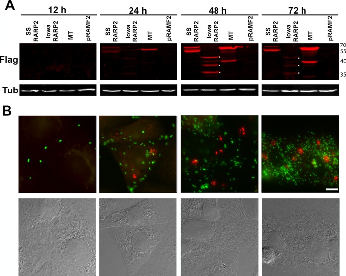
FIG 3 .
Expression of RARP-2 during infection. (A) FLAG-tagged SS-RARP-2, Iowa-RARP-2, MTase, and pRAMF2 empty vector control were overexpressed from R. rickettsii Iowa in Vero76 cells (MOI of 1) and sampled at 12, 24, 48, and 72 hpi for immunoblotting with an anti-FLAG antibody. Dots to the right of the bands indicate the major breakdown products of Iowa-RARP-2 at 48 and 72 hpi. Reduced signal at 72 h is presumably due to further proteolysis of the products. Tubulin was used as a loading control. (B) Cultures infected with SS-RARP-2-FLAG were fixed at times corresponding to panel A and stained with anti-FLAG antibodies (red) for observation by immunofluorescence assay. GFP-expressing rickettsiae are green. Bar, 10 µm. Corresponding Nomarski differential interference contrast images are provided.

