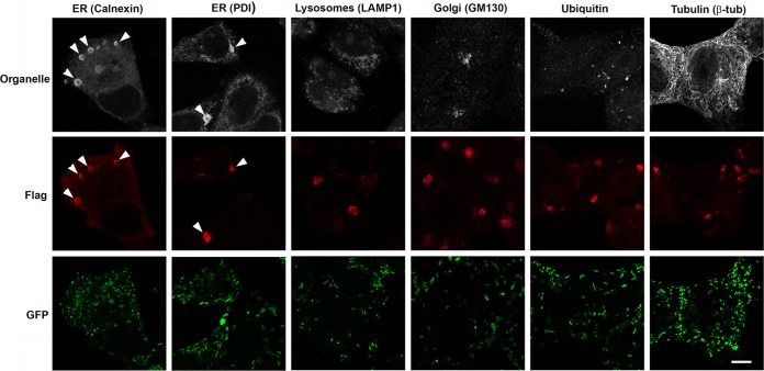
FIG 5 .
RARP-2 structures colocalize with ER markers. R. rickettsii SS-RARP-2 was expressed from R. rickettsii Iowa in Vero76 cells (MOI of 1). Cultures were stained with anti-FLAG antibody (red) and observed by immunofluorescence assay after 48 hpi. Pleomorphic structures were observed in the cytosol of infected cells. Cultures were counterstained for various organelles: ER, lysosomes, Golgi apparatus, ubiquitin, and β-tubulin (white). RARP-2 structures colocalize only with the ER markers calnexin and PDI. Rickettsiae expressing GFP are green. Arrowheads identify multiple RARP-2 vesicular structures colocalizing with ER markers. Bar, 10 µm.

