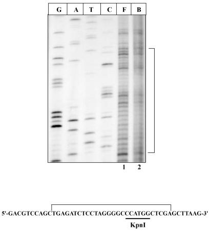
Figure 5.
Footprinting of KpnI methyltransferase. Free (F) and bound (B) lanes contain pUC18 and pACMK plasmids, respectively, after treatment with (OP)2Cu and extension with DNA polymerase I (Klenow) as described in Materials and Methods. The bracket indicates the protected region both in the figure and in the sequence of the multiple cloning site of pUC18 plasmid. The recognition sequence for KpnI methyltransferase is underlined. G, A, T and C are the sequencing lanes.

