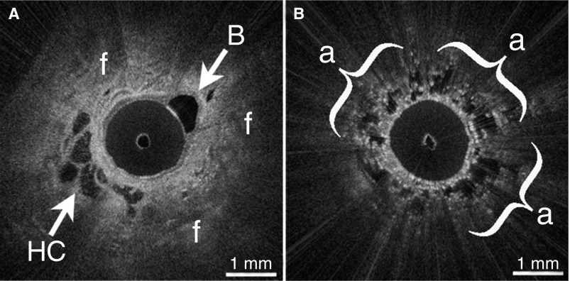
Figure 2.
(A) In vivo endobronchial optical coherence tomography (OCT) identified microscopic honeycombing (HC) and destructive peripheral fibrosis (f) in a patient with nondiagnostic high-resolution computed tomography and indeterminate lung biopsy. A branching bronchiole (B) is also visible. (B) OCT visualized spatial heterogeneity as regions of preserved alveoli (a) adjacent to fibrosis.

