Fig. 2.
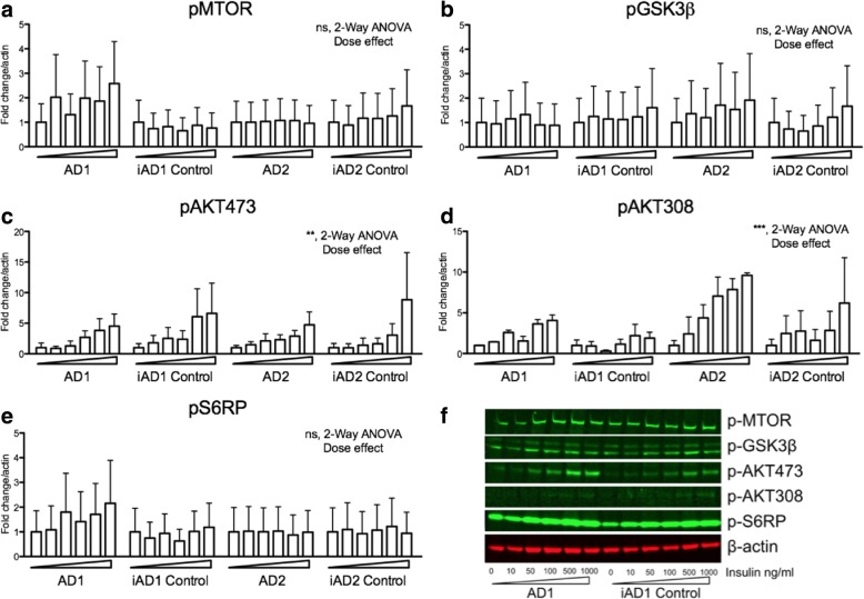
Downstream insulin signaling pathway molecules display similar status of phosphorylation in iPSC-derived wildtype and PSEN2N141I basal forebrain cholinergic neurons (BFCNs). a-e Western blot analysis of iPSC-derived BFCNs. Cells (DIV 34) were insulin deprived overnight before the addition of insulin at 0, 10, 50, 100, 500, 1000 ng/ml. Cells were collected after a 30-min exposure. Quantifications of western blot data is normalized over actin and expressed as fold change to vehicle treated cells. These data correspond to results of two to three independent experiments. Dose dependency was detected by 2-way ANOVA where indicated. ** P < 0.01; *** P < .001. f Representative blots from a single experiment showing three of the lines

