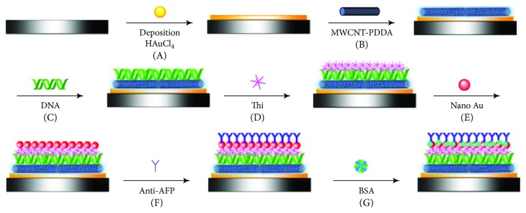
Figure 5.
Schematic view of the fabrication process of an immunosensor reported by Ran et al. (A) Deposition of Au nanoparticles. (B) Coating of MWNTs-PDDA layer. (C) Immobilization of DNA film. (D) Formation of thionine layer. (E) Assembly of gold nanoparticles. (F) Anti-AFP loading. (G) BSA blocking (reprinted from Ran et al. [127] with permission).

