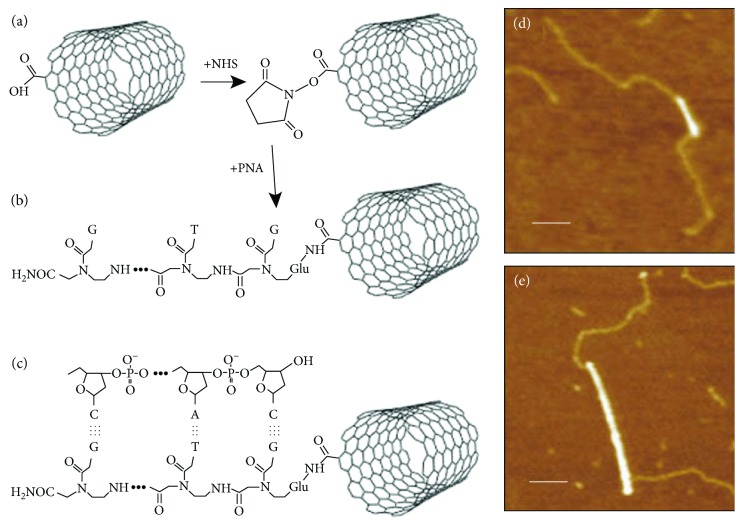
Figure 9.
Hybridization of PNA attached SWNTs and DNA. (a and b) PNA was attached to an end of SWNT with N-hydroxysuccinimide (NHS) esters. (c) Hybridization of DNA with PNA attached SWNT. (d and e) AFM images of PNA-SWNTs. Bright lines indicate SWNTs. The paler strands represent bound DNA. Scale bars are 100 nm. Diameters of SWNTs were 0.9 nm and 1.6 nm in (d) and (e), respectively (reprinted from Williams et al. [263] with permission).

