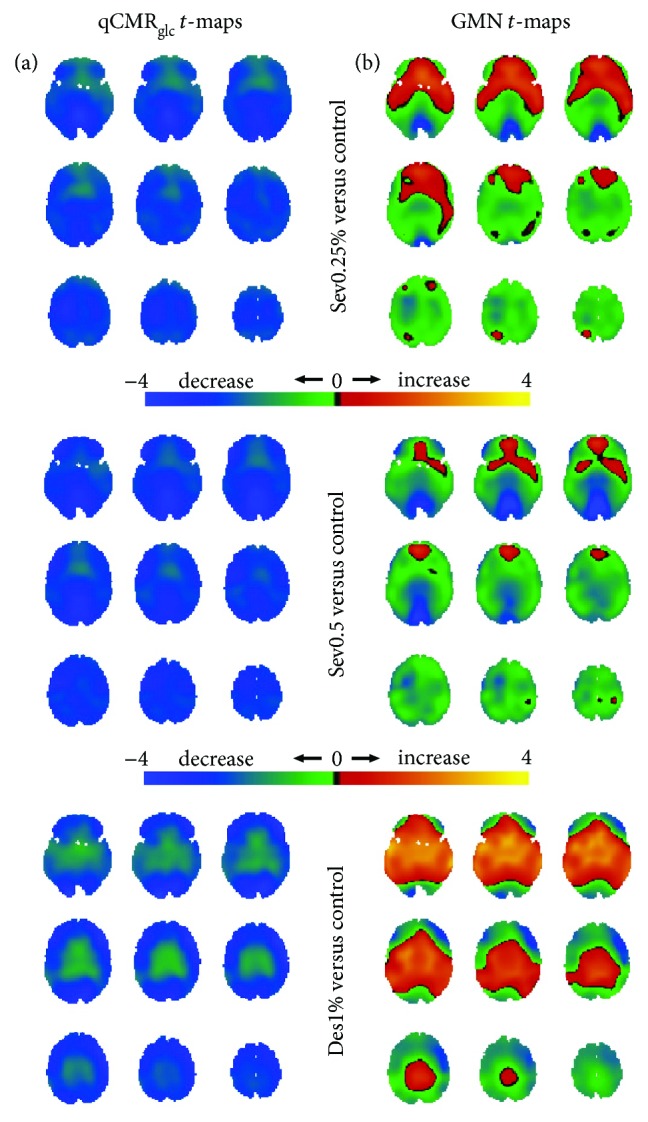
Figure 4.

Spatial distributions of metabolic variations with sedation (i.e., Des1%, Sev0.25%, and Sev0.5% in Figure 2(b)) versus the control group (i.e., HAEC in Figure 2(a)), shown with respect to unthresholded Student's t-maps using (a) qCMRglc images and (b) GMN images. (a) For Des1%, Sev0.25%, and Sev0.5% groups, the unthresholded t-maps with qCMRglc indicated globally unidirectional metabolic decreases with sedation. (b) However, the unthresholded t-maps with GMN depicted regions with metabolic increases and decreases upon sedation. Based on validation of qCMRglc to aCMRglc-HYD (Figures 1 and 2; Table 2), without GMN the global decreases corresponded to about 0.05 μmol/g/min (Des1% ≈ Sev0.5% < Sev1%) and with GMN the deemphasis on global changes put the focus on the regional differences. See Figure S4 for thresholded maps (Table 4).
