Figure 5.

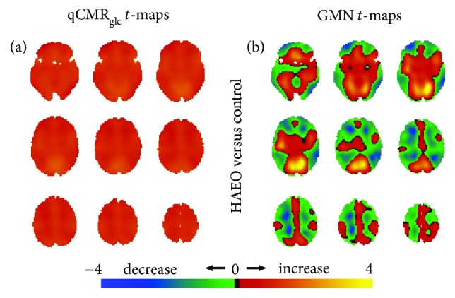
Spatial distributions of metabolic variations with healthy participants with eyes open (i.e., HAEO in Figure 2(a)) versus the eyes closed control group (i.e., HAEC in Figure 2(a)), shown with respect to unthresholded Student's t-maps using (a) qCMRglc images and (b) GMN images. (a) For the HAEO group, the unthresholded t-maps with qCMRglc indicated the presence of globally unidirectional metabolic increases with eyes open. (b) Conversely, the unthresholded t-maps with GMN revealed regions of increased and decreased metabolism with eyes open. Based on validation of qCMRglc to aCMRglc-HYD (Figures 1 and 2; Table 2), without GMN the global increases corresponded to about 0.05 μmol/g/min while with GMN, the global changes were minute. See Figure S5 for thresholded maps (Table 4).
