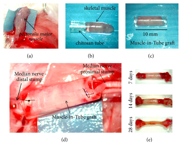
Figure 1.

Preparation of the muscle-in-tube graft. A longitudinal piece of the pectoralis major muscle was removed (a) and used to fill the chitosan conduit (b), to obtain a 10 mm long muscle-in-tube graft (c). The graft was then used to repair a median nerve defect (d). Pictures showing the regenerated nerves at different time points postsurgery (e). The conduit has been removed and a suture was used to mark the proximal stump (on the right in the pictures).
