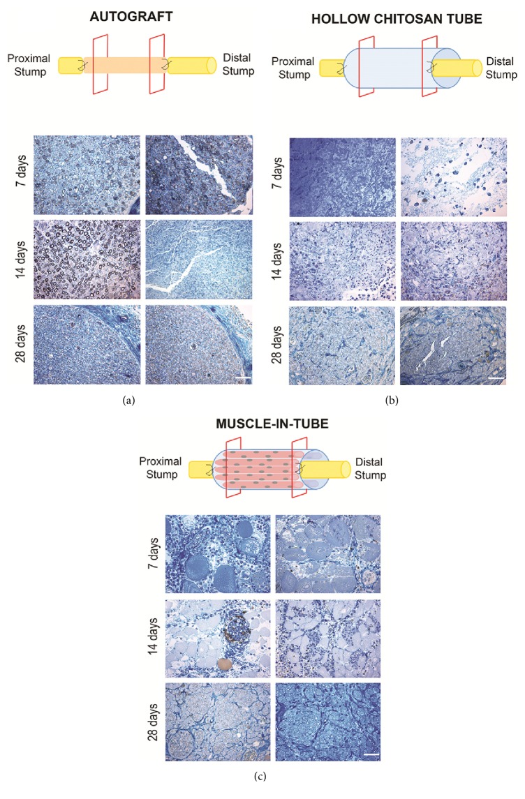
Figure 3.
Representative high resolution light images of toluidine-blue stained semithin transverse sections of regenerated median nerve repaired with autograft (a), hollow chitosan tube (b), or muscle-in-tube graft (c). Images were taken inside the grafts, both proximally (left columns, about 1,5 mm from the proximal suture point) and distally (right columns, about 1,5 mm from the distal suture point), at different time points (7, 14, 28 days after repair). Bar: 40 μm.

