
Figure 7.
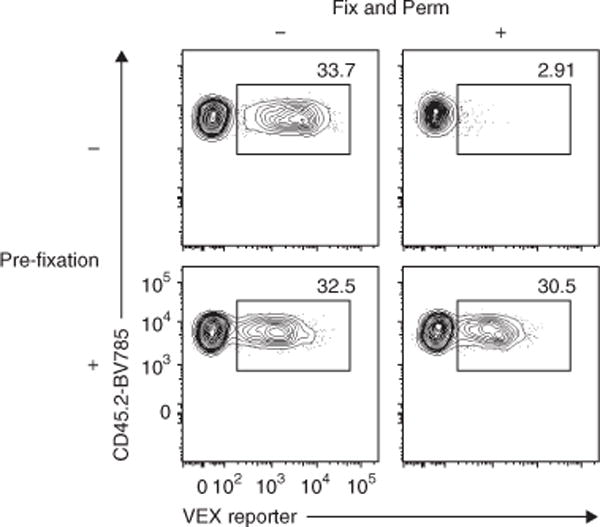
Pre-fixation of RV-transduced T cells prevents the loss of RV-derived marker detection. Spleens containing empty-VEX+ RV-transduced wild-type P14 cells were harvested on day 36 after Arm infection. After staining with surface CD8, TCR Vα2, CD44, CD45.1 and CD45.2, spleen cells were treated with mock PBS or 2% (vol/vol) PFA in PBS at 4 °C for 20 min. Then half of the cells were fixed and permeabilized (Fix and Perm) using an eBioscience Foxp3 staining kit according to the manufacturer’s instructions. The remaining half of the samples were kept in PBS at 4 °C during the Fix and Perm steps. All samples were analyzed immediately after Fix and Perm procedures. Representative flow plots gated on P14 cells are shown. Numbers in the plots indicate percentage of VEX+ cells among total P14 cells. Data are representative of three independent experiments (1–2 technical replicate(s) per condition in each experiment). All animal experiments depicted in Figure 7 were performed in accordance with the institutional animal care and use guidelines of the University of Pennsylvania.