Figure 1.
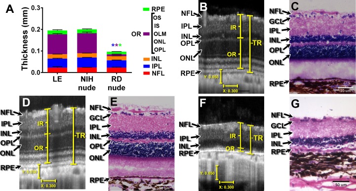
SD-OCT and histology of LE, NIH, nude, and RD nude rats. (A) Quantification of retinal layer thickness using OCT scans: LE rats (n = 6) and NIH nude rats (n = 6) were compared to RD nude rats (n = 2) at the age of 5 to 7 months. Asterisks correspond to P < 0.05; color indicates significant difference for corresponding layer color. (B, D, F) Sample images of cross-sectional retina OCT scans in (B) LE rat (age 6.8 months), (D) NIH nude rat (age 6.8 months), and (F) RD nude rat (age 4 months). Vertical bar: 50 μm; horizontal bar: 300 μm. (C, E, G) Hematoxylin and eosin staining of the retina for (C) LE rat (age 7 months), (E) NIH rat (4 months), and (G) RD rat (6.6 months). Scale bars: 50 μm. RPE, retinal pigment epithelium; OS, outer segments; IS, inner segments; OLM, outer limiting membrane; ONL, outer nuclear layer; OPL, outer plexiform layer; INL, inner nuclear layer; IPL, inner plexiform layer; GCL, ganglion cell layer; NFL, nerve fiber layer; OR, outer retina (OPL, ONL, OLM, IS, OS); IR, inner retina (NFL, GCL, IPL, INL); TR, total retina (from nerve fiber layer to RPE).

