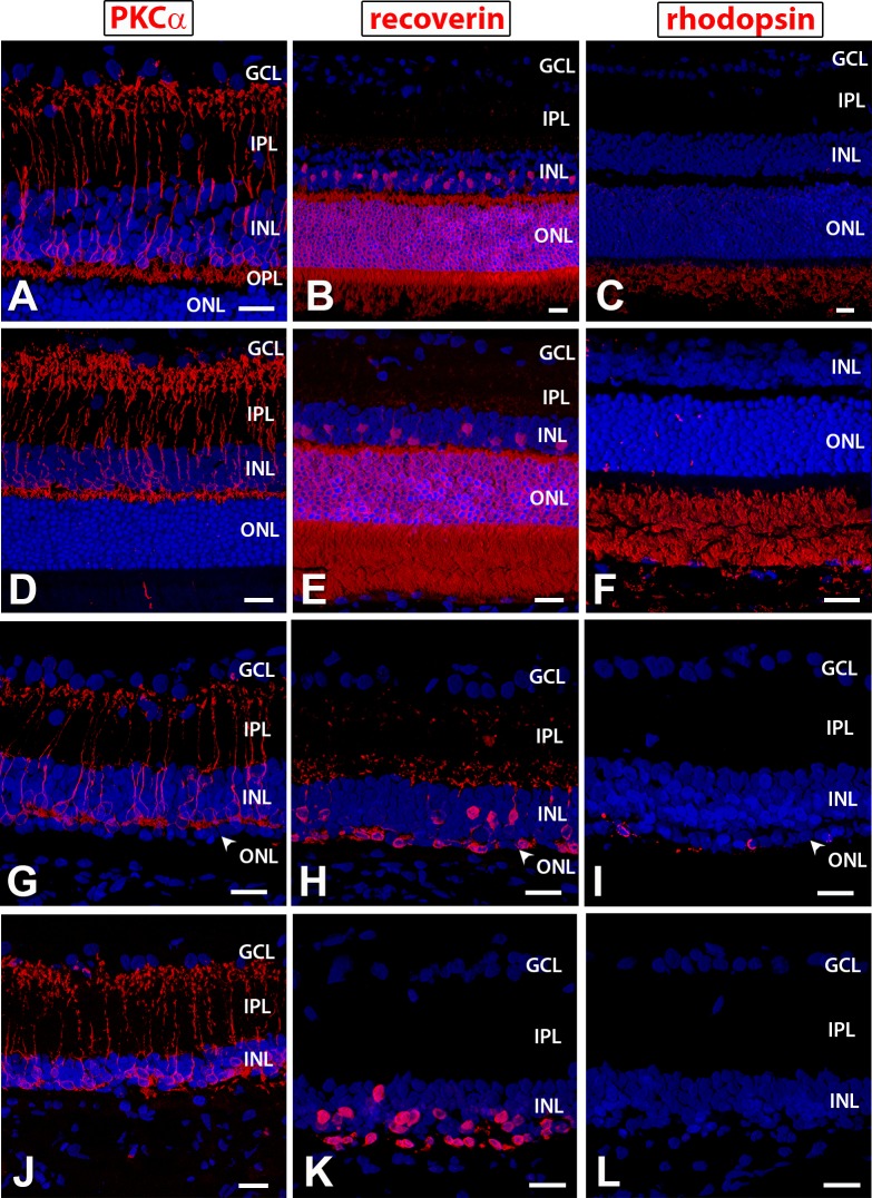
Figure 2.
Immunohistochemistry on nonsurgery controls for PKCα, a marker of rod bipolar cells (A, D, G, J); recoverin, a marker for cone bipolar cells and photoreceptors (B, E, H, K); and rhodopsin (C, F, I, L). (A–C) 1-month-old NIH nude rat retina (section somewhat obliquely cut so layers appear much thicker than with the 4-month-old NIH retina); (D–F) 4-month-old NIH nude rat retina; (G–I) 1-month-old RD nude rat retina: (G) outer plexiform layer still clearly recognizable with PKC staining; (H) the outer nuclear layer is reduced to one cell layer; (I) few cells in the ONL are immunoreactive for rhodopsin; (J–L) 6.6-month-old RD nude rat retina: few photoreceptors left. 3D images of confocal ×40 stacks. Scale bars: 20 μm. GCL, ganglion cell layer; IPL, inner plexiform layer; INL, inner nuclear layer; OPL, outer plexiform layer; ONL, outer nuclear layer.

