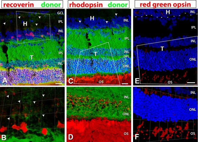
Figure 8.
hPAP (donor, green), recoverin (red), rhodopsin (red), and red–green opsin staining in well-laminated transplants. Nuclei in (A, C, E, F) are labeled in blue. Arrowheads point to transplant processes in the host IPL. (A, C, E) are 3D images of confocal stacks; Scale bars: 20 μm. (B, D, F) are zoomed in and rotated 3D opacity images rendered in Volocity. (A, B) recoverin (photoreceptor and cone bipolar cells); (A) shows laminated structure of transplant with cone bipolar cells evident in the INL of transplant and host. (C, D) Rhodopsin (red: rods). (E, F) red–green opsin (red: red–green cones); the dashed line in (E) represents the transplant border. H, host; T, transplant; GCL, ganglion cell layer; IPL, inner plexiform layer; INL, inner nuclear layer; ONL, outer nuclear layer; OS, outer segments.

