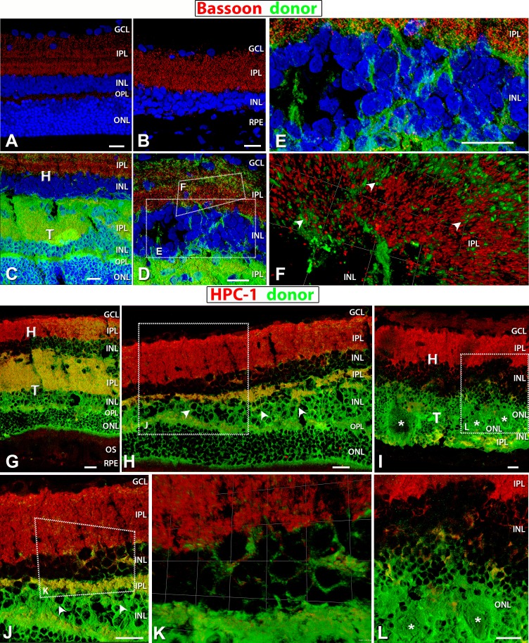
Figure 10.
Staining for Bassoon (red: ribbon synapses) (A–F) and HPC-1 (red: syntaxin, membranes of amacrine cell bodies and plexiform layers) (G–L). Donor cells are labeled green (hPAP) and nuclei in blue (DAPI). (A) Bassoon labeling of the IPL and OPL in a 4-month-old normal NIH nude retina. (B) The remaining IPL ribbon synapses labeled with Bassoon in a 6.6-month-old RD retina with sham surgery (no. 14). No OPL recognizable. (C–E) Ribbon synapses present in the host and donor IPL in laminated transplant no. 5. (D) Enlargement at the host–transplant interface with donor processes (green) infiltrating host. Areas of magnification in (E) and (F) are indicated. (E) Transplant processes (green) evident in the host INL and IPL. (F) 3D opacity image of donor processes (green) extending into the host retina IPL with colocalization of red and green indicating synaptic contact between the two (arrowheads). (G, H, J, K) Laminated transplant no. 5 with HPC-1 staining evident in the host and transplant IPL, and (H, J) amacrine cells in transplant INL (arrowheads). (K) Enlarged 3D opacity image at the host–transplant interface with colocalization of red and green staining indicating transplant processes making synaptic contact with host. (I, L) Strong HPC-1 labeling of host IPL and very little in donor within rosetted (white asterisks) transplant no. 1 IPL, but amacrine cells are present. (L) Enlargement of a small area with few transplant processes extending into host. Scale bars: 20 μm.

