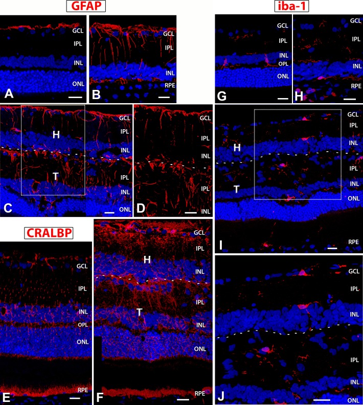
Figure 12.
GFAP, CRALBP, and Iba1 expression in NIH nude rats, sham RD, and surgery RD rats. In images of transplant 5 (C, D, F, I, J), a dashed line indicates the approximate border between transplant and host. (A) NIH nude retina (7.5 months): GFAP expression is restricted to nerve fiber layer astrocytes. (B) Sham RD rat (6.6 months) and (C, D) transplant 5 (rat age 6.9 months) show similar GFAP expression levels in activated Müller cells, but (D) is a zoom of a region in (C) that demonstrates there is only a partial glial barrier at the transplant–host interface. The host and transplant retina contains radial processes of Müller glial cells that also wrap around blood vessels. (E) CRALBP localization in NIH nude rat (3.9 months): stain of RPE and radial processes and cell bodies of Müller cells. (F) Transplant 5: strong CRALBP staining of host and transplant radial Müller glia and host RPE. Partial limiting membrane between transplant and host recognizable in (F). (G–J) Microglia marker Iba1; (G) NIH nude retina (7.5 months): The resident microglia are localized in the inner retina (IPL, INL, OPL) and are few in number. They appear more amoeboid and are likely in surveillance mode. A sham RD rat (age 6.9 months) (H) and transplant 5 (I, J) show increased Iba+ expression and branching morphology indicative of microglial activation and recruitment. (J) is an enlargement of the boxed area in image (I) demonstrating process extension and activation. 3D images of confocal stacks. Scale bars: 20 μm.

