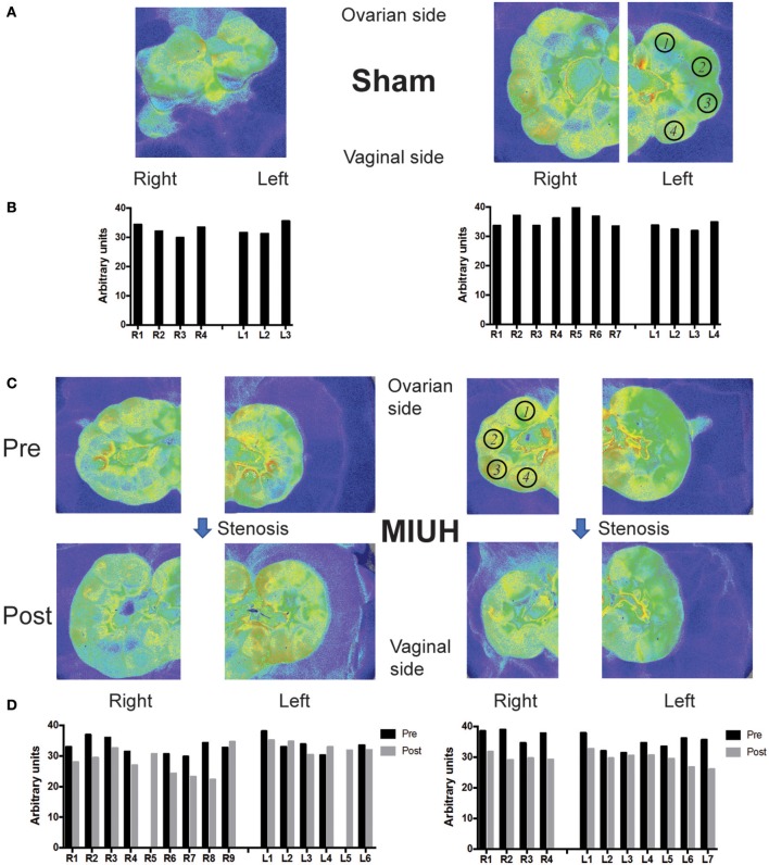
Figure 1.
Laser speckle flowmetry and blood flow measures in the fetuses and placentas of pregnant rats that experienced either sham operation or microcoil stenosis at embryonic day 17 (E17) leading to mild intrauterine hypoperfusion (MIUH). (A) Images of laser speckle of the uterine horns in two representative sham rats at E17. (B) Quantification of blood flow in fetuses that correspond to the laser speckle images in (A). (C) Images of laser speckle showing changes in uterine blood flow before (Pre) and 1 h after microcoil stenosis (Post) at both sides of the ovarian artery. (D) Blood flow changes during coils stenosis in fetuses in both dams shown in (C). The blood flow decreased 1 h after stenosis (Post) compared to that before stenosis (Pre) in most of the fetuses and placentas (not shown). The location of pups in the right (R) and left (L) horns of the uterus is denoted from 1 to the maximal number of pups in each side, with 1 at the closest location to the ovary and the last one at the closest location to the vagina. The region of interest (ROI) for blood flow measures of the fetuses is depicted in (A,C) by black open circles, with numbers that correspond to the locations of the fetus on each uterus horn.

