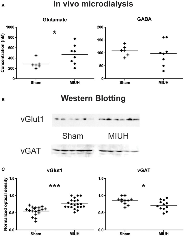
Figure 4.
Long term effects of MIUH on the excitation/inhibition balance in the hind limb area of the sensorimotor cortex. (A) Plots of glutamate and GABA extracellular levels assessed by in vivo microdialysis in adult rats. Compared to sham rats, MIUH induced a significant increase in glutamate release (U = 8.0; p < 0.04) while the release in GABA did not differ between the two groups (U = 18.0; p = 0.4, n.s.). (B) Typical immunoblots for vGlut1, vesicular transporter of glutamate, and vGAT, vesicular transporter of GABA in the two groups of rats. (C) Plots of glutamate and GABA intracellular levels assessed by Western blots of vGlut1 and vGAT, respectively. Note that both measures indicate an increase in glutamate levels [t(1, 37) = 4.8; p < 0.0001] while GABA levels were reduced [t(1, 21) = 2.4; p < 0.02] after MIUH, compared to sham rats. *p < 0.05; ***p < 0.001.

