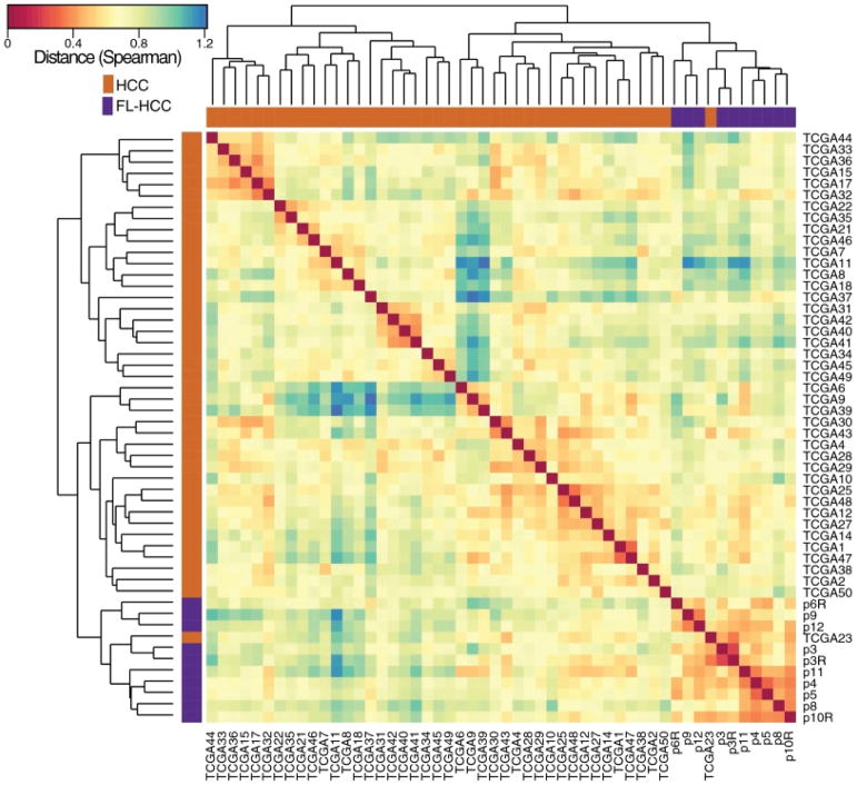
Figure 2. FLC and HCC have distinct molecular characteristics.
The transcriptome of fibrolamellar tumor relative to the adjacent normal and hepatocellular carcinoma tumor, relative to the adjacent normal, were analyzed and characterized by hierarchical clustering (complete method) with a spearman rank correlation matrix for all 3708 protein coding genes that passed the DESeq2 expression filter (|log 2 fold change| > 1, FDR < 0.01, and ranked by moderated t statistic ranking). The transcriptome of the fibrolamellar samples (purple) segregated away from those of hepatocellular carcinoma (orange) [Data shown is for 10 FLC and 42 HCC from over 400 HCC patients analyzed]. An example is shown of one patient out of five found that had been reported to be HCC, but the transcriptome segregated with FLC. This particular example was from a young adolescent who was diagnosed with HCC without a molecular characterization. An analysis showed the presence of a fusion between the genes DNAJB1 and PRKACA consistent with this patient actually having fibrolamellar (see figure 3). A histological sample was not available in this specific case. Figure from (10).

