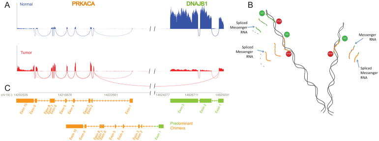
Figure 3. Molecular basis for FLC: A deletion in a chromosome 19 produces a chimeric gene.
Figure A. The reads for the mRNA were mapped onto the genome. An increased number of reads for PRKACA, the catalytic subunit of protein kinase A, in the tumor sample (red) relative to the normal (blue) in exon 2 through 10, and in the reads spanning the exons, demonstrated the increased expression of this gene. In addition in the tumor there were reads that spanned between the start of exon 2 of PRKACA and the end of exon 1 of DNAJB1, a member of the heat shock protein family. Figure B. This pattern of reads demonstrate that in the tumor there is the normal transcript for DNAJB1 (green), and the normal transcript for PRKACA (goldenrod) and a chimeric transcript with the first exon of DNAJB1 and the 2nd to final exons of PRKACA. This chimeric transcript was confirmed by Sanger sequencing (28). Figure C. The formation of the chimeric transcripts, while maintaining the normal transcripts, is consistent with a deletion of ~400 kB in one copy of chromosome 19. This deletion was confirmed by Sanger sequencing (28). Figure adapted from (28).

