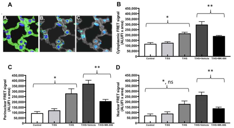
Figure 2. Subcellular localization of FRET signal intensity.
A) Pulmonary localization of ALOX5 and ALOX5AP in Trauma and Hemorrhagic Shock-induced lung injury. Quantification and the intracellular location of ALOX5 and ALOX5AP are shown. The cellular structures were partitioned into (A) cytosolic (B) perinuclear and (C) nuclear domains that were mutually exclusive (illustrated in these representative images in green). Nuclei are blue and the cytosolic domains are gray (B and C).
B) The molecular association between ALOX5 and ALOX5AP in the cytoplasm increases after T/HS when compared to Control and T/SS (*, p<0.05). FRET intensity significantly decreased after administration of MK-886 when compared T/HS+Vehicle (**, p<0.01).
C) The molecular association between ALOX5 and ALOX5AP in the perinuclear domain increases after T/HS when compared to Control and T/SS (*, p<0.01). FRET intensity significantly decreased after administration of MK-886 when compared T/HS+Vehicle (**, p<0.01).
D) The molecular association between ALOX5 and ALOX5AP in the nuclear domain increases after T/HS when compared to Control (*, p<0.01) but not T/SS (p>0.05). FRET intensity significantly decreased after administration of MK-886 when compared T/HS+Vehicle (**, p<0.01).
ALUFI, arbitrary linear units of fluorescence intensity.

