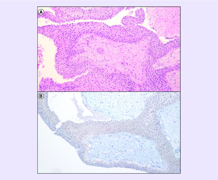
Figure 1. . Pathology findings.
H&E-stained section revealed a well-circumscribed neoplasm with well-differentiated squamous epithelium surrounding a hypocellular fibrous stroma with stellate and bipolar-shaped cells (A: 100×). No mitotic figures, atypia or pleomorphism was seen. Immunohistochemistry for mutated BRAF-V600E was positive (B: 100×, Ventana VE1 clone).
H&E: Hematoxylin and eosin; VE1: BRAF-V600E.

