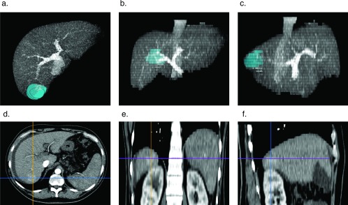
Figure 1.
3D MIP reconstruction of tumor rendered as a volume for surgical planning. The tumor is rendered as a volume within reconstructed 3D MIP DICOM of the liver in the venous phase: axial (A), coronal (B), and right sagittal (C) views. DICOMs were read with the Horos open-source medical image viewer.11 Axial (D), coronal (E), and right sagittal (F) arterial phase images are of a segment 7 hepatoma.

