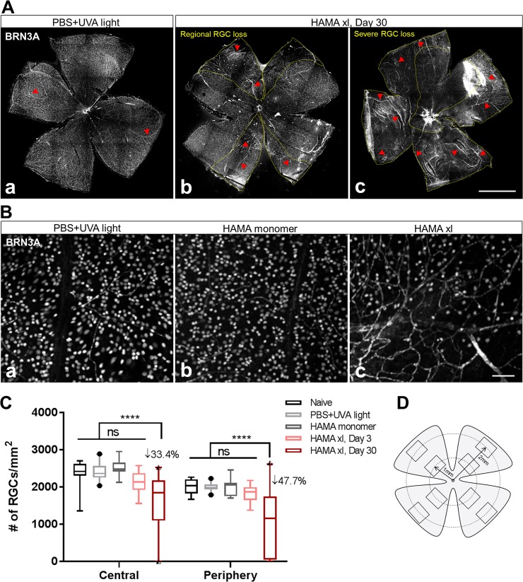
Fig 2. Ocular hypertension induced by HAMA xl led to significant loss of retinal ganglion cells (RGCs) one month post-operation.
(A) Representative graphs of the Day 30 retinal flatmounts immunostained with BRN3A, a RGC nucleus marker. Loss of RGCs was observed from the hypertensive eyes treated with HAMA xl on Day 30. Note: the mouse monoclonal BRN3A antibody used in the present study cross-reacted with the blood vessels (red arrowheads) in the retina which became more visible in the degenerative retinas. Yellow dotted lines in b, c delineate the regions with more prominent RGC loss. Scale bar: 1 mm. (B) Representative micrographs of BRN3A immunolabeling of RGCs from normotensive (a. PBS+UVA light; b. HAMA monomer) and hypertensive (c. HAMA xl) retinas. Scale bar: 50 μm. (C) Quantification of RGC density based on BRN3A+ nuclei count from retinal flatmounts. Graph was shown as interleaved box & whiskers with 95% confidence interval. n = 11–12 eyes/group for naive, HAMA monomer (Day 30) and HAMA xl (Day 3) groups; n = 20 eyes/group for PBS+UVA light (Day 30) and HAMA xl (Day 30) groups, **** P<0.0001, ns: non-significant. Two-way ANOVA followed by Tukey’s multiple comparisons test. (D) Schematic indicating the sampling of eight 563μm x 422μm rectangle area in the retinal flatmount from four quadrants at two eccentricities (central vs periphery) from the optic nerve head (ONH) for RGC quantification (refer to S1 Fig for technical details).

