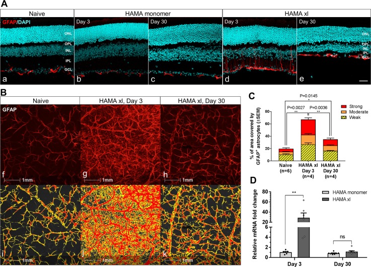
Fig 5. Astrocyte activation was observed in the hypertensive retinas with more prominent reactivity detected on Day 3 compared to Day 30.
(A) Immunostaining of GFAP in retinal vertical sections. HAMA xl induced dramatic astrocyte activation with higher reactivity detected at early time point Day 3 (d) compared to Day 30 (e). In contrast, PBS+ UVA light (data not shown) or HAMA monomer (b,c) did not induce detectable astrocyte activation compared to naive controls. Images were collected from approximately 1.2–1.5mm from the optic nerve head in the retinal vertical sections. Scale bars: 50 μm. (B) Representative GFAP-immunoreactivity in retinal flatmounts. Top panel: GFAP immunostianing in retinal flatmounts from naive (f), hypertensive retina on day 3 (g) and day 30 (h); bottom panel: signals delineated by HALO software for corresponding images in f,g,h. No activation of astrocytes was noted in the retinal flatmounts from the PBS+UVA or HAMA monomer controls, data not shown. (C) Quantification of GFAP-immunoreactivity by HALO based on the total area covered by GFAP-immunofluorescence and the signal intensity. The intensity of GFAP-immunofluorescent signal was categorized as strong, moderate and weak by HALO. GFAP-immunoreactivity was significantly upregulated in the hypertensive retinas on Day 3 (P = 0.0027) and Day 30 (P = 0.0145) compared to naive controls. A significant attenuation of GFAP-immunoreactivity was also detected on Day 30 compared to Day 3 (P = 0.0036). Error bars: SEM. (D) GFAP mRNA was significantly upregulated in the hypertensive retinas on Day 3, which attenuated largely on Day 30. ** P = 0.008, multiple t-tests. Error bars: SEM.

