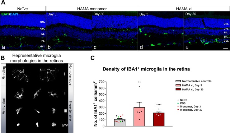
Fig 6. Ocular hypertension led to significant microgliosis in the hypertensive retinas.
(A) Immunostaining of IBA1 to label microglial cells in the retinal vertical sections from normotensive (naive and HAMA monomer controls) and hypertensive (HAMA xl) retinas. Activation of microglia was observed in the hypertensive retinas on both Day 3 (d) and Day 30 (e). PBS+ UVA light (data not shown) or HAMA monomer (b,c) did not induce detectable microglia activation. Scale bar: 50 μm. (B) Representative microglia morphologies in the normotensive and hypertensive retinas. (C) Quantification of IBA1+ cells revealed significant increase of microglia cells in the hypertensive retinas on Day 3 and Day 30. One-way ANOVA. ** P = 0.0014, *** P<0.0001, error bars indicate SEM.

